Urinary system
The urinary bladder (Fig. 2) lies in the lower part of the abdominal cavity behind the pubic bone. It is a hollow organ surrounded by a layer of smooth muscles. Its function is first to collect the urine coming from the two ureters and then to lead the urine through the urethra for excretion.
The urinary bladder is closed by a special closing mechanism – the sphincter muscle/pelvic floor muscles – so that no urine is lost before the usually voluntary emptying process.

Structure and function of the urinary system
The urinary system consists of the kidneys and the urinary tract, which includes the renal pelvis, ureter, urinary bladder and urethra. The function is the same in men and women, but there are anatomical differences.
The function of the kidneys is to filter out the body's metabolic waste products that are dissolved in the blood and to filter water from the bloodstream. The resulting urine is passed through the renal pelvis and ureters into the urinary bladder (Fig. 1).
The antidiuretic hormone (a hormone that inhibits the excretion of water, known as ADH or vasopressin) is of decisive importance in urine production. It significantly reduces the amount of urine by means of water recovery in the kidneys. At night the level of antidiuretic hormone rises in the body, thereby leading to reduced urine production during sleep. Normally, this means that we do not have to go to the toilet during the night.

Control of bladder emptying
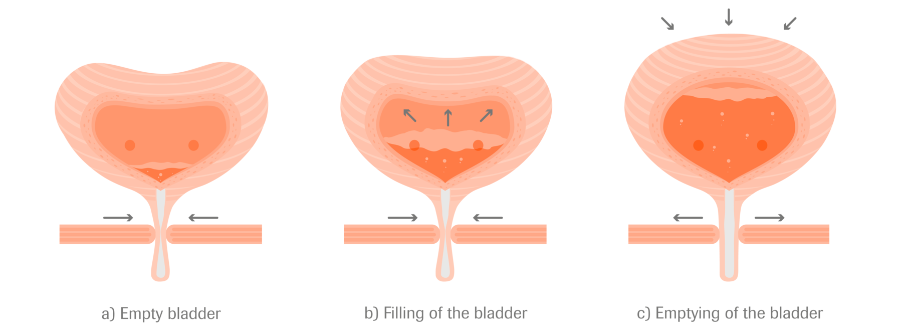
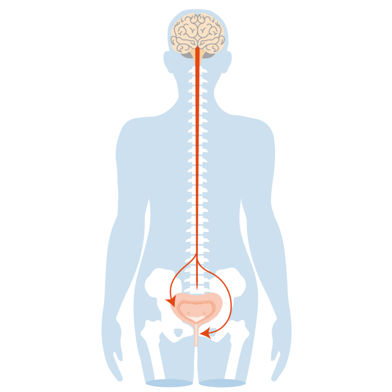
When empty (Fig. 3a), the bladder muscle is relaxed and the sphincter muscle tensed, the bladder is closed. During filling (Fig. 3b), the bladder muscle remains relaxed and the sphincter muscle remains tensed, the bladder is closed. During bladder filling, the nerves in the bladder wall constantly register the filling level and report it to the brain (Fig. 4).
Once the bladder is full, the nerves send a message to the brain that emptying must take place. Emptying can be controlled voluntarily. During emptying (Fig. 3c), the sphincter muscle relaxes, i.e., the bladder outlet opens and the urine can flow out. At the same time, the bladder muscle is tensed so that the urine is also squeezed out of the bladder. After emptying is completed, the sphincter muscle tightens again, the urethra is closed again and the bladder muscle relaxes (Fig. 3a).
The function of the kidneys and the urinary system are identical in men and women. However, there are differences in anatomy below the bladder – essentially determined by the sex organs.
In women, the urethra is only 3–4 cm long and ends in the vulval vestibule. As a result of this and the proximity of the urethra to the anus, women are generally more susceptible to urinary tract infections than men. In women, the pelvic floor, which is part of the bladder closure mechanism, is additionally perforated by the birth canal (vagina). Vaginal births or a prolapse of the uterus can lead to damage to the pelvic floor and thus to the closure mechanism of the bladder. During pregnancy, additional pressure can be exerted on the urinary bladder or its position can be changed, while malfunctions in bladder emptying are also possible.
In men, the urethra has a length of about 20 cm and ends at the tip of the glans on the penis. Directly below the bladder the prostate is located. If the prostate is enlarged, it can affect the bladder's closure mechanism. This can make emptying more difficult – even if the urinary system is otherwise intact.